Ball Cages for Automated Microscope XY Stages
An automated microscope scans a tissue slide tile by tile. The X-Y carriage moves between tiles. The stitch between adjacent tiles is off. At the edge of the field, the image is slightly soft, as though the focus plane tilted as the stage reached the side of travel. The objective is centered, the condenser is aligned, and the scan pattern is correct. The carriage guide has gone soft.
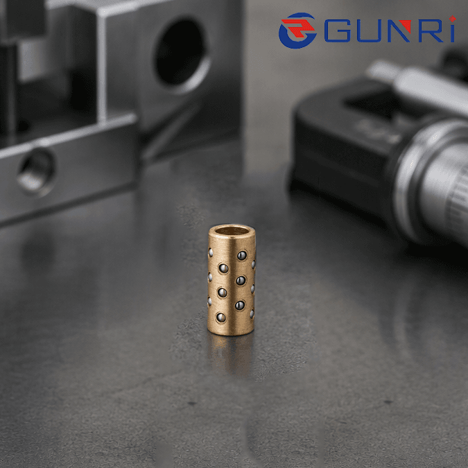
Motorized microscope stages are optical instruments as much as they are motion systems. A small amount of lateral play, a small tilt of the carriage, or a sticky Z focus axis shows up directly in the image. A precision ball cage is the internal part that keeps the carriage and the focus spindle moving on their intended axes.
What fails on a scanning stage
A microscope XY stage carries a slide or a well plate across a field. The carriage rides on some form of linear guide — crossed rollers, recirculating ball rails, or a ball-caged dovetail. The Z axis moves the objective or the nosepiece up and down for focus. Both need to be straight, smooth, and repeatable.
When a stage ages, three things tend to happen. The carriage develops side play, so the tile grid drifts. The Z focus spindle binds at one end of travel, so auto-focus hunts. The fine-adjustment micrometer has a dead band, so manual focusing feels mushy. Each of these traces back to how the moving element is guided, and in many sub-assemblies that guidance is a ball cage.
Where the cage sits in the stage
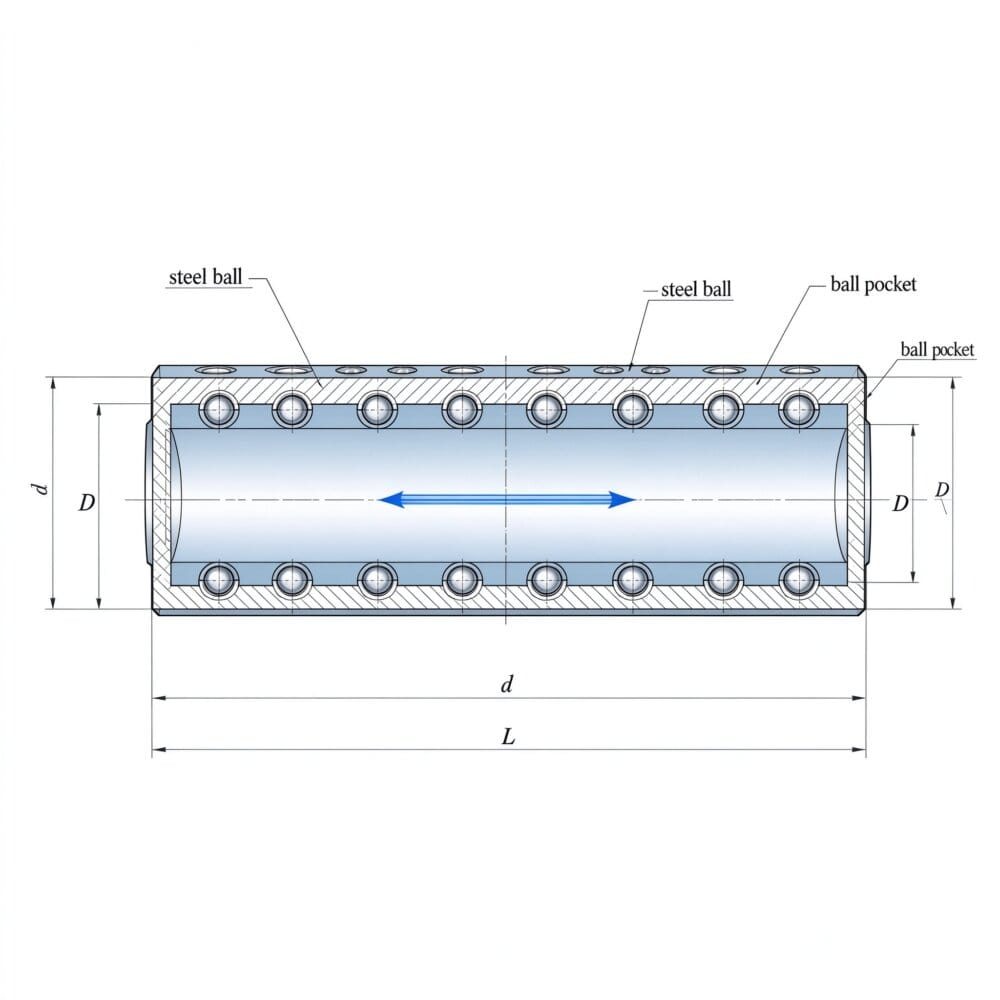
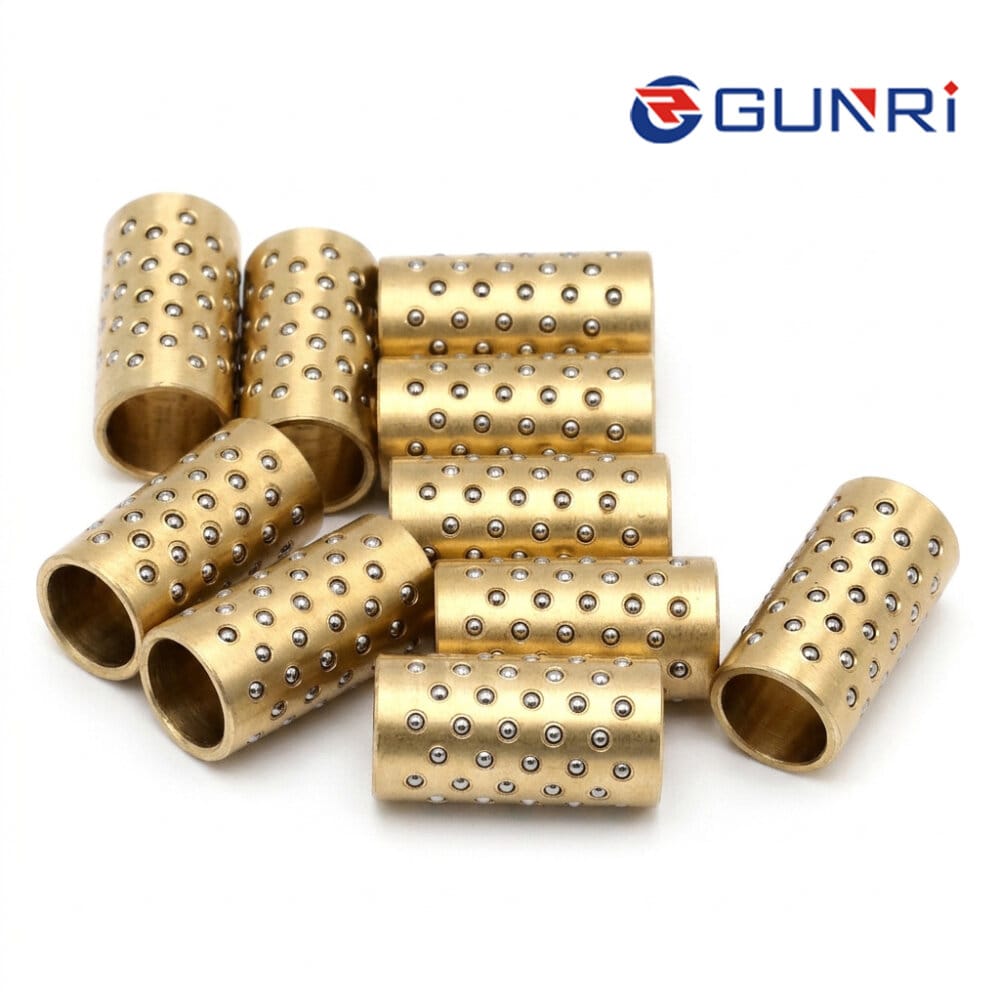
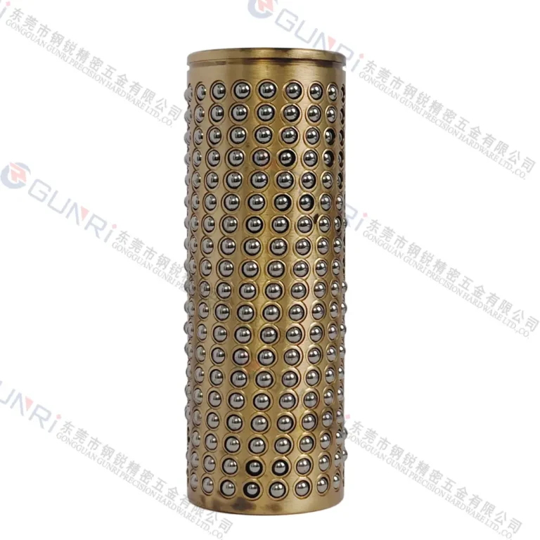
A ball cage on a microscope stage is usually a thin ring of captured balls between a cylindrical plunger and a housing bore. It shows up in three places:
- The Z focus spindle. The objective turret or nosepiece rides up and down on a spindle. A cage around the spindle keeps it from cocking as it moves, so focus comes in at the same center every time.
- The fine-adjustment plunger. The coarse/fine focus knob pushes a small plunger against a spring. A cage around that plunger removes side play and tightens the fine focus.
- The XY carriage cross-slide. On smaller manual or motorized stages, the moving carriage can ride on ball-caged ways instead of a full rail system. The balls are held in a slotted retainer that follows the carriage through its stroke.
Why slotted windows matter here
Stage travel is finite — a few tens of millimeters in X, similar in Y, and a few millimeters in Z. If the balls are held in short round pockets, the cage itself has to shift back and forth inside the housing as the carriage moves. That shift introduces a small relative motion between cage and carriage, and a small side load at the ends of travel.
Slotted windows let the balls travel with the carriage. The balls stay under the carriage through the whole stroke, and the cage stays fixed in the housing. We cover this geometry at slotted window ball cages. It is the same approach used on optical translation stages, where a micrometer-driven carriage has to move cleanly through its full range.
Multi-row arrangements for the nosepiece
A rotating nosepiece carrying multiple objectives is heavy and off-axis. A single ball row lets it tilt. Two rows, separated along the spindle, create a longer guide span and resist the moment. We write about the layout at multi-row ball cages. Where the stage moves quickly between tiles, an aluminum cage keeps the moving mass down, so the motor can settle fast between scans.
Sizing and inspection
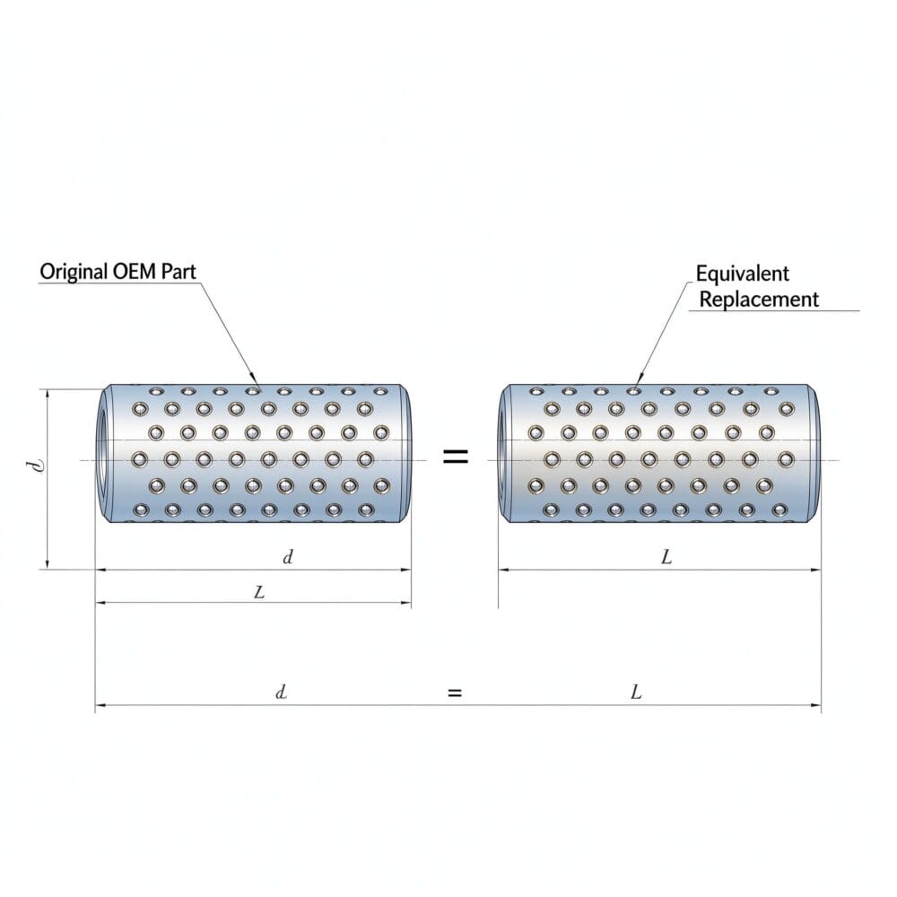
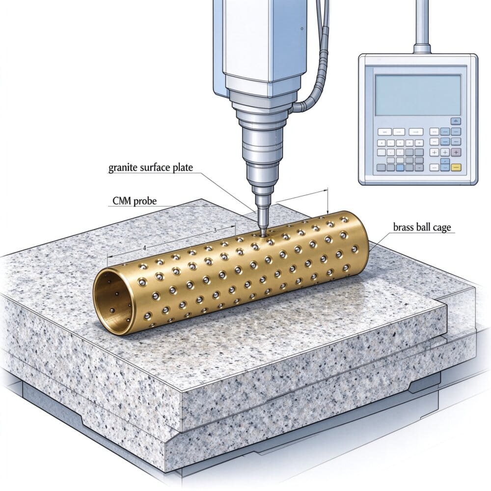
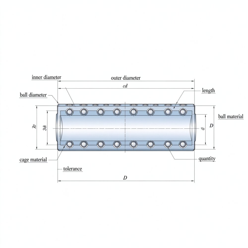
Microscope sub-assemblies are compact. We build cages down to a 2.0 mm ID with 0.6 mm balls and an 8 mm minimum length. Larger research microscopes and stage controllers scale up. Every cage is CMM-inspected on the ball circle and the critical diameters. We accept STEP, IGES, and SLDPRT drawings, and we hold no MOQ, so a prototype focus spindle can be validated before a production run.
What to look for during teardown
When a scanning stage shows a soft stitch, pull the carriage and inspect the ball row. A ball that has dropped out of its pocket leaves the carriage supported on three points instead of a circle, and that shows up as a slight rotation of the slide as it reaches the end of travel. A bright, grooved band on the spindle means the balls have been running dry. A discolored or flattened ball means the preload was too high and the balls are brinelling.
Each of these is a cage problem, not an optics problem. Replacing the cage restores the geometry; re-aligning the optics will not.
If your scanning stage has developed a soft stitch or a sticky focus axis, the internal ball row is worth a look. Send the drawing of the current guide and we will quote a staked replacement.
We build custom cages from prototype through production. Start on the contact page with your drawing or sample, and we will work out the ball arrangement, material, and staking style with you.